Helium-based magnetometers
A glimpse of Hardware and Physics
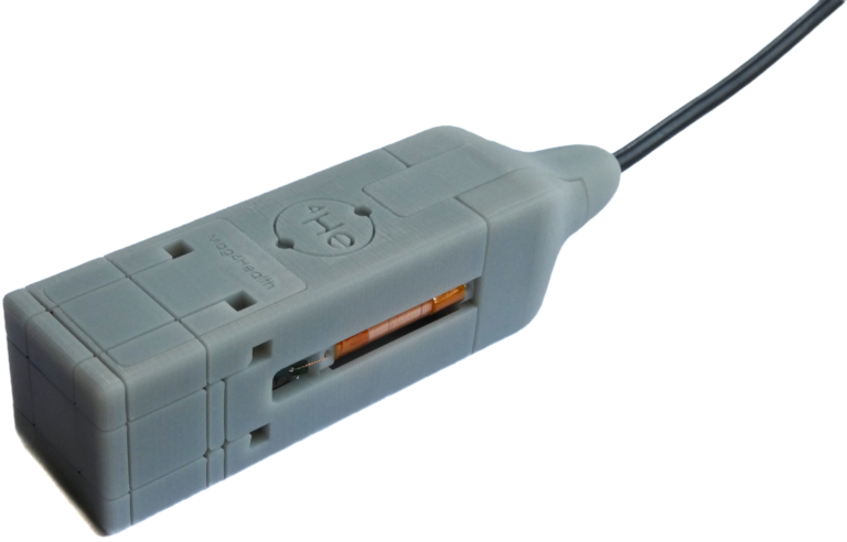
In the MAG4Health sensor the sensitive element is a gas of Helium (4He) atoms in their 23S1 metastable state. Helium is a noble gas, very stable over time, and has demonstrated its robustness and lifetime in Space magnetometry.
The size of the cell containing the 4He gas atoms is cylindrical with a 1 cm internal diameter and 1 cm internal height. The bottom of the sensor is surrounded by small 3-axis Helmholtz coils, which are used to apply both the RF fields and the compensation fields.
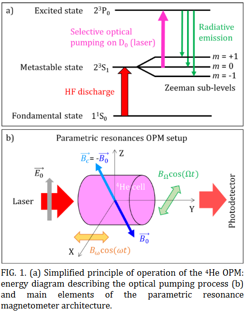
- First, we apply a high frequency (HF) discharge that brings 4He atoms from the ground state to their 23S1 metastable state which is a spin-one state, sensitive to the magnetic field.
- Then a alignment polarization is created by optically pumping the metastable state at 1083.2 nm wavelength with linearly polarized light.
- Two radio-frequency fields at different frequencies (9 and 40 kHz) allow exciting parametric resonances. The measurement of the three components of the magnetic field 𝐵0⃗⃗⃗⃗ is obtained from the measurement of the compensation currents.
More details on our sensors can be found in our 2021 paper (Fourcault et al., https://doi.org/10.1364/OE.420031)